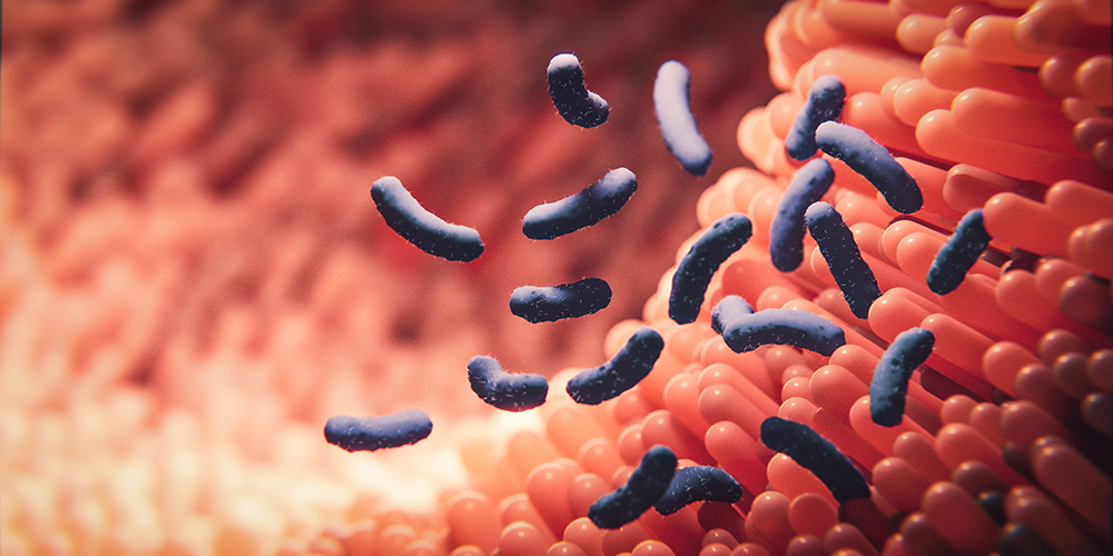
Nanosized bacterial speargun
One micrometer long and less than thirty nanometers wide – more than thousand times thinner than a human hair – that’s the size of a bacterial “speargun”. This so-called type VI secretion system is an almost unbeatable weapon in fighting against competitors or invading host cells. In a recent publication in “Cell” the team of Prof. Marek Basler at the Biozentrum of the University of Basel shed light on the structure of this secretion apparatus at atomic level.
27 February 2015
Many types of bacteria including pathogens causing cholera or pneumonia use the type VI secretion system (T6SS) to kill eukaryotic host cells or surrounding competitors to enhance their survival. This versatile tool plays an important role not only in virulence but also in symbiosis and interactions between bacteria. A team of researchers led by Marek Basler, Professor at the Biozentrum, University of Basel, has now resolved the structure of an important component of the T6SS at the atomic level and provides new insights into the assembly of this nanomachine.
Atomic details of a bacterial nanomachine
The T6SS injection apparatus can be imagined as a very tiny speargun that allows bacteria to inject toxic proteins directly into target cells. This nanomachine was discovered almost ten years ago but details of its structure were still unknown. Marek Basler’s group applied cryo-electron microscopy and state-of-the-art imaging available at C-CINA and newly developed computer technologies to build an atomic model of a specific T6SS component – the so-called sheath.
“The sheath is a sophisticated tube that has the amazing ability to contract in less than five milliseconds to push a toxic spear out of a cell”, says Basler. “We could now show at atomic resolution how the single subunits of the contracted sheath are connected to form a cog-wheel like ring and how these rings connect to form a very long tube. Inside of the sheath the subunits are linked via a so-called handshake domain which is critical for assembly and contraction”, explains Basler.
Contractile sheath similar in viruses
Moreover, structural comparisons between the T6SS sheath of the cholera pathogen and sheath of bacteriophages – viruses that attack bacteria – revealed that both injection systems evolved from a common ancestor. However, bacteria evolved a distinct outer layer to allow for a quick recycling of the individual sheath components. “Our study provides new deep insights into the structure and the basic mechanism how this nanomachine works”, says Basler. “In ongoing experiments we want to uncover the assembly of the extended sheath and its transition to the contracted structure.” This knowledge may serve as the basis for the development of novel antibacterial therapies or development of new macromolecule delivery systems.
Original article
Mikhail Kudryashev, Ray Yu-Ruei Wang, Maximilian Brackmann, Sebastian Scherer, Timm Maier, David Baker, Frank DiMaio, Henning Stahlberg, Edward H. Egelman, Marek Basler.
Structure of the type VI secretion system contractile sheath
Cell, published online 26 February 2015. doi:10.1016/j.cell.2015.01.037
Further information
Prof. Marek Basler, University of Basel, Biozentrum, phone +41 61 267 21 10, email: marek.basler@unibas.ch


