
Cartilage and bone development: three paths to skeleton formation
In vertebrates, the skeleton of different regions of the body arises from different precursor cells. Researchers at the University of Basel have now discovered that these skeletal cells do not just differ in their developmental origin, but also in their gene regulation – which may be a key to the vertebrates’ evolutionary success story.
27 March 2025 | Angelika Jacobs
From the skull to the smallest bone in your pinky toe, the skeleton acts as internal scaffolding to give stability to the body, and forms protective cocoons around important organs. Despite their similar structure, however, not all bones are created equal: in vertebrates (including humans), the various parts of the skeleton arise from different groups of precursor cells during embryonic development. During this process, each group produces its own set of regulator proteins and goes through its own developmental program to produce cartilage and bone. Researchers from the University of Basel have reported these findings in the scientific journal Nature Communications.
Three construction teams, each with their own blueprint
One type of precursor cell forms the skull and facial bones, another the spinal column and ribs, and a third type the skeleton of the limbs. “You can imagine it as three construction teams, each building one story of a house,” explains Professor Patrick Tschopp of the Department of Environmental Sciences at the University of Basel. “The three teams start with different materials, blueprints and tools, but you end up with three stories that are structurally and functionally similar.”
The skull and facial bones arise from what are known as neural crest cells, which are produced on the back of the embryo and are developmentally closest to the cells of the central nervous system. The precursor cells of the spine and ribs are somitic mesoderm cells, which arise from the sides of the embryo’s back and form – besides bones – also muscles and parts of the skin. The third group originates from the lateral plate mesoderm on the embryo’s flanks, and goes on to form the skeleton of the arms and legs, along with parts of the ribcage.
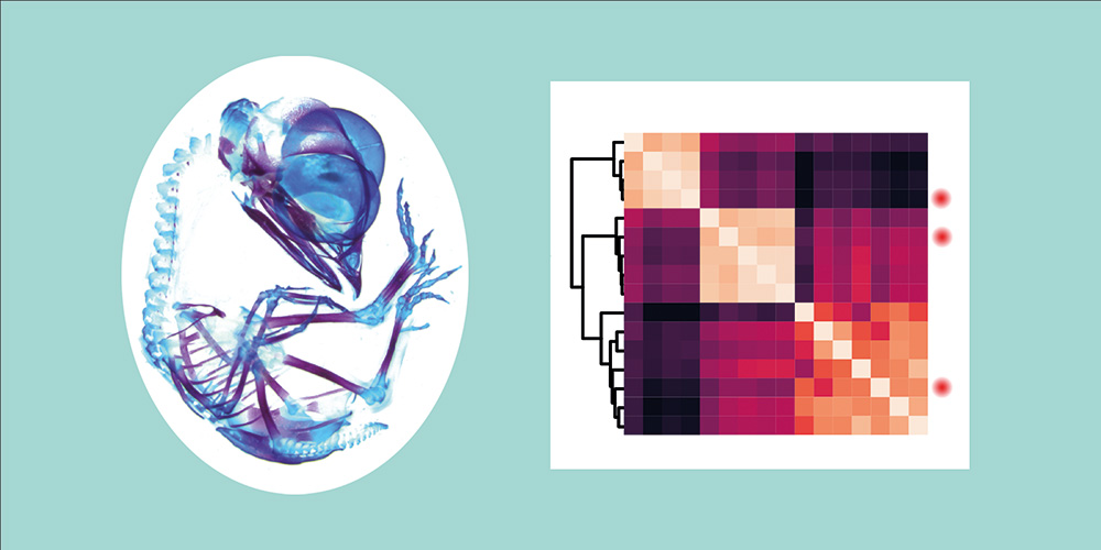
By carrying out single cell-based analyses in chicken embryos, the research team discovered that the three groups of cells all use different regulatory mechanisms to drive the developmental program that creates skeletal cells. “From these results, we conclude that skeletal cells in the different regions of the body are actually not as alike as was previously thought,” says bioinformatician Dr. Menghan Wang, one of the two lead authors of the study. “Rather, they appear to be different cell types involved in the production of a similar tissue,” says developmental biologist Dr. Ana Di Pietro-Torres, the second lead author.
Why these differences are an advantage
What seems unnecessarily complicated at first glance might actually be one of the keys to the vertebrates’ evolutionary success: “If the skeleton of different regions of the body is determined by different blueprints, these parts of the skeleton can also change independently from one another,” says Patrick Tschopp. “That might explain why vertebrates have evolved so many different types of skeletons.”
Original publication
Menghan Wang, Ana Di Pietro-Torres et al.
Distinct Gene Regulatory Dynamics Drive Skeletogenic Cell Fate Convergence During Vertebrate Embryogenesis
Nature Communications (2025), doi: 10.1038/s41467-025-57480-8
